

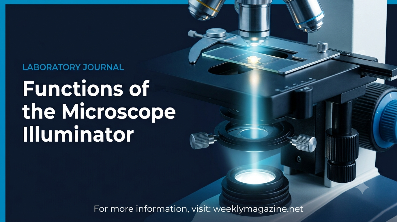
7 Essential Functions of the Illuminator on Microscope: Complete Guide
The primary function of the illuminator on microscope systems is to produce a steady, high-intensity, and controllable light beam that penetrates or reflects off a specimen to make microscopic structures visible to the human eye.
Types: LED, Halogen, Mirror & UV
Impact: Image Contrast & Clarity
Even the most expensive objective lenses and eyepieces are virtually useless without a reliable, bright light source. In biological and materials research, understanding the exact function of the illuminator on microscope instruments allows technicians, researchers, and students to obtain high-resolution, glare-free, and high-contrast images. Located within the base or mounted externally, the illuminator serves as the energy foundation of the optical path.
In this comprehensive analytical report on weeklymagazine.net, we examine the fundamental mechanisms of microscope lighting, compare legacy and modern light sources, clarify the differences between condensers and illuminators, and explain how the key function of the illuminator on microscope hardware controls specimen visibility under high magnification.
What Is the Function of the Illuminator on Microscope Equipment?
The primary function of the illuminator on microscope systems is to provide consistent, evenly distributed light to pass through or reflect off the specimen being observed. Positioned typically in the base of compound light microscopes, its job is to generate photons that travel through the iris diaphragm and condenser lens directly to the objective optics.
- Primary Objective: Provides controlled illumination necessary for magnifying translucent or opaque samples.
- Common Light Types: Modern LEDs, tungsten-halogen bulbs, fluorescent lamps, or adjustable concave mirrors.
- System Synergy: Works directly with the condenser (which focuses light) and the diaphragm (which adjusts aperture size).
1. The Fundamental Role of Light in Optical Microscopy
Microscopes magnify objects using light waves. When a specimen is mounted on a glass slide, human eyes cannot perceive details unless light rays pass through the tissue or reflect off its surface into the objective lens. The main function of the illuminator on microscope units is to serve as the originating light engine for this optical journey.
Without an active light source, a specimen appears entirely dark or blurred under high magnification objectives (such as 40x or 100x oil immersion lenses). High magnification lenses possess narrow numerical apertures, meaning the primary function of the illuminator on microscope tools is to demand intense light rays to create a crisp, well-defined focal image.
2. 7 Key Functions of the Illuminator on a Microscope
To fully grasp how light microscopy operates, it helps to break down the specific tasks performed by the illuminator light assembly during sample inspection:
- Generating Controlled Photons: The core function of the illuminator on microscope devices is emitting steady light waves toward the stage, eliminating reliance on unpredictable ambient room light.
- Providing High-Intensity Output for Magnification: Higher objective magnification reduces the light reaching the eye; the illuminator compensates by boosting light output.
- Ensuring Uniform Field Aperture Lighting: Distributes light evenly across the target area to prevent uneven shadows or dark corners (vignetting).
- Enabling Transmitted vs. Reflected Observation: Drives light directly through thin, biological tissues (transmitted light) or bounces light off solid surfaces (reflected/episcopic light).
- Regulating Variable Rheostat Intensity: Modern illuminators include voltage controls or dimmers so operators can adjust brightness based on specimen transparency.
- Supporting Phase Contrast and Darkfield Modes: Delivers bright, aligned light rays necessary for specialized filtering techniques.
- Establishing Köhler Illumination Balance: Works alongside the field diaphragm to create uniform lighting across the specimen without showing the lamp filament in the final image.
3. Types of Microscope Illuminators Compared
Microscope manufacturing has transitioned from simple physical mirrors to advanced solid-state light-emitting diodes (LEDs). Each light source type exhibits distinct operational characteristics, though the primary function of the illuminator on microscope systems remains identical across models:
| Illuminator Light Type | Color Temperature & Spectrum | Primary Advantages & Common Uses |
|---|---|---|
| LED (Light Emitting Diode) | Daylight Cool White (~6000K) | Energy efficient, long lifespan (>50,000 hrs), generates zero heat, ideal for living biological cells. |
| Tungsten / Halogen Bulb | Warm Yellowish Spectrum (~3200K) | Very bright output, excellent color rendering for stained histology slides; requires blue daylight filters. |
| Fluorescent / Mercury Vapor | Ultraviolet & High Energy Blue Wavelengths | Excites fluorophores in molecular biology, clinical diagnostics, and genetic research. |
| Pivoted Mirror (Legacy) | Relies on ambient room lighting | Requires no electricity; commonly found on simple vintage or portable field educational microscopes. |
Explore our primary editorial desk on WeeklyMagazine Main Portal for expert technology reviews. For complementary academic, biological, and analytical educational breakdowns, check out The Better Readers Educational Hub.
4. What is the Difference Between an Illuminator, Condenser, and Iris Diaphragm?
A common area of confusion among microscopy students is differentiating between the illuminating parts under the microscope stage. While they work together, each component has a distinct role:
- The Illuminator (Light Source): Generates light rays. The essential function of the illuminator on microscope bases is acting as the raw source of photon emission.
- The Condenser Lens Assembly: Positioned directly above the illuminator and beneath the stage, it gathers diffuse light rays from the illuminator and focuses them into a tight cone directed at the specimen.
- The Iris Diaphragm: Mounted inside or under the condenser, it controls the numerical diameter of the light cone, adjusting image contrast and depth of field.
5. How to Optimize Microscope Illumination for Clear Images
To achieve maximum image clarity and avoid eye strain during extended laboratory sessions, follow these standard operational guidelines:
- Start at Low Rheostat Output: Turn on the illuminator at low voltage to prevent sudden light bursts that can cause eye discomfort or blow out bulb filaments.
- Align the Condenser Height: Adjust the condenser position so light covers the entire field of view without leaving dark edges.
- Adjust Brightness as Magnification Increases: When shifting from a 4x objective to a 100x oil immersion objective, gradually increase the illuminator brightness to maintain image clarity.
- Clean the Glass Lens Filter Regularly: Dust on the protective illuminator glass lens creates shadows in captured photographs; keep it clean with lint-free lens paper.
6. Frequently Asked Questions (FAQs & PAA)
Q1: What is the primary function of the illuminator on a microscope?
The primary function of the illuminator is to provide a steady, bright, and uniform source of light that illuminates the specimen, making it clearly visible through the magnifying optics.
Q2: What is the purpose of an illuminator in simple terms?
In simple terms, an illuminator acts as the light bulb or light mirror of the microscope, supplying necessary light so user eyes or digital cameras can view sample details clearly.
Q3: Are the illuminator and light source the same thing in a microscope?
Yes, “illuminator” and “light source” are interchangeable terms in microscopy. Both refer to the component—whether an LED, halogen bulb, or mirror—that supplies light to the system.
Q4: What is the function of the condenser and illuminator on a microscope?
The illuminator produces light rays, while the condenser gathers those diffuse rays and focuses them into a narrow, intense light beam targeted directly onto the glass slide specimen.
Q5: Why do high-power objective lenses require brighter illuminator settings?
High-power objective lenses (such as 40x, 60x, and 100x) have smaller lens apertures that collect less total light. Increasing illuminator output maintains clarity and prevents dark, grainy images.
7. Final Remarks on Modern Microscope Light Technology
Mastering optical microscopy starts with understanding how light is generated and controlled. The core function of the illuminator on microscope instruments goes beyond simply casting light—it forms the energy foundation that determines image contrast, color accuracy, and overall resolution across all scientific investigations.
Syeda
Syeda is an expert SEO specialist, content strategist, and link builder writing for weeklymagazine.net. She specializes in crafting comprehensive educational content, technical equipment analyses, and search-optimized scientific guides.